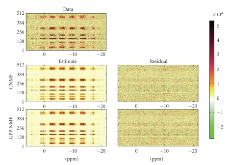
Figure 4.
Brain imaging data matrix (top) along with the estimated decomposition and residual for the CNMF (middle) and GPP-NMF (bottom) methods. In this view, the results of the two decompositions are very similar, the data appears to be modeled equally well and the residuals are similar in magnitude.

