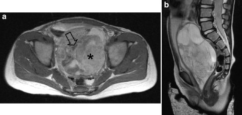
Fig. 11.
A 2-year-old girl presented with a mass in the vagina. a Axial T1-W contrast-enhanced image shows the mass with heterogeneous enhancement. The tumour has both solid (asterisk) and fluid (open arrow) compartments. b Sagittal T2-W MR image shows the mass with mixed signal intensity. The bladder is displaced anteriorly and the uterus cannot be visualized. Histopathology: embryonal RMS

