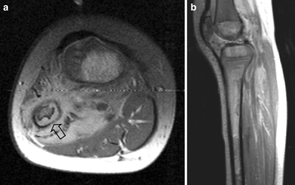
Fig. 12.
A 4-year-old girl presenting with a mass in the left lower leg. a Axial T1-W contrast-enhanced MR image shows an ill-defined mass circumferential to the fibula. Note the cortical thinning (open arrow) of the fibula. b Sagittal PD-weighted image shows diffuse bone marrow metastases. Histopathology: embryonal RMS

