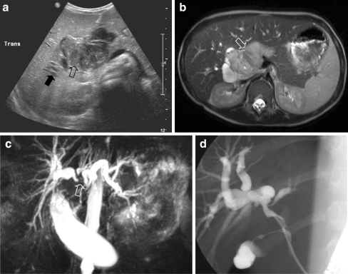
Fig. 16.
An 8-year-old boy presented with abdominal pain and jaundice. a US image shows a central process in the liver hilum (open arrow) and dilatation of the intrahepatic bile ducts (solid arrow). b T2-W MR image shows a circumscribed lesion with increased signal intensity (open arrow). c MRCP image shows intrahepatic bile duct dilatation. Note that the right and left duct systems do not communicate (open arrow). d ERCP image (ERCP performed in order to insert a stent in the common bile duct). Histopathology: embryonal RMS

