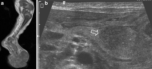
Fig. 18.
A 4-day-old girl born with a lump on the left foot. Antenatal ultrasonography at 20 weeks showed no abnormalities. a T1-W MR image shows a large inhomogeneous mass arising from the left foot. b Abdominal US image shows popliteal and inguinal nodal invasion, and hepatic and pancreatic metastases (open arrow). Due to the poor prognosis, no therapy was given, and the child died several weeks later. Histopathology: poorly differentiated soft-tissue sarcoma without distinct translocations

