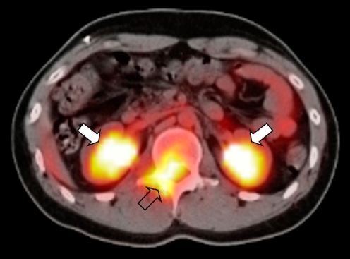
Fig. 8.
A 19-year-old boy with a history of treated metastatic RMS presented with low back pain. The PET-CT image shows intense 18F-FDG uptake in the spinal canal (open arrow). Physiological excretion of the radiopharmaceutical via the kidneys is visible (solid arrows). Histopathology: embryonal RMS

