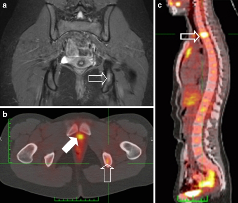
Fig. 9.
Two years after initial diagnosis the patient shown in Fig. 6 presented at the outpatient clinic complaining of back pain. a Coronal STIR image of the pelvis shows discrete increased signal intensity in the left ischium (open arrow). b Subsequently acquired PET-CT image confirms the presence of recurrent disease in the same location (open arrow). Note excretion of tracer into the urinary bladder (solid arrow). c PET-CT image also shows a second lesion in the thoracic spine (open arrow). Additional rib and pleural metastases were also visible (not visible on this image)

