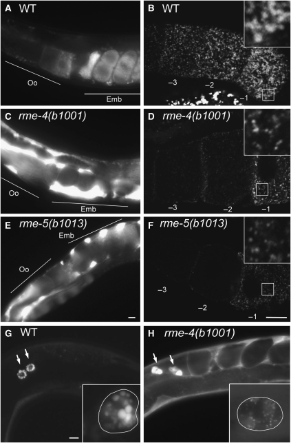
Figure 1.
rme-4 and rme-5 mutants display endocytosis defects. (A, C, E) YP170–GFP endocytosis by oocytes of adult hermaphrodites. In wild-type, YP170–GFP is efficiently endocytosed by oocytes (A). In the rme-4(b1001) and rme-5(b1013) mutants, endocytosis of YP170–GFP by oocytes is greatly reduced and YP170–GFP accumulation in the body cavity is greatly increased (C, E). Positions of oocytes (Oo) and embryos (Emb) are indicated. (B, D, F) Gonads from wild-type or mutants expressing YP170–GFP were dissected and fixed before imaging. In the oocytes of mutant animals, the overall intensity of YP170–GFP fluorescence was reduced, and YP170–GFP that was internalized was observed in abnormally small vesicles. Insets show enlargements ( × 3) of the boxed area. Oocytes proximal to the spermatheca are numbered as −1. (G, H) Coelomocyte endocytosis. GFP secreted from body-wall muscle cells (ssGFP) is taken up by coelomocytes and accumulates in endosomes/lysosomes of wild-type coelomocytes (G). In rme-4(b1001), increased accumulation of ssGFP in the body cavity was observed, indicating poor endocytosis by coelomocytes (H). Arrows indicate each coelomocyte. Enlarged images of coelomocytes are shown in the insets. Cell boundaries of coelomocytes are outlined for clarity. Bars, 10 μm.

