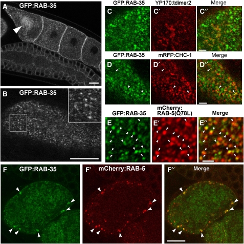
Figure 4.
Subcellular localization of rescuing GFP–RAB-35 in the germ line. (A, B) GFP–RAB-35 was expressed under germline-specific promoter control and observed in live animals. Middle (A) and top (B) focal planes are shown. In developing oocytes, GFP–RAB-35 mainly localizes to small puncta in the cortex. Localization of GFP–RAB-35 to the yolk granules deeper in the cytoplasm becomes prominent in oocytes close to the spermatheca (an arrowhead in panel A). An enlarged ( × 2) image of the boxed area is shown in the inset (B). Bars, 10 μm. (C–C″, D–D″, E–E″) Colocalization of GFP–RAB-35 with endocytic markers in oocytes. GFP–RAB-35 and either YP170–tdimer2 (yolk protein; C–C″), mRFP–CHC-1 (D–D″) or mRFPCherry–RAB-5(Q78L) (E–E″) were co-expressed in the germ line and their subcellular localization was observed in live animals. Bars, 2 μm. (F–F″) Colocalization of GFP–RAB-35 and mRFPCherry–RAB-5(WT) in one-cell-stage embryos. Bar, 10 μm.

