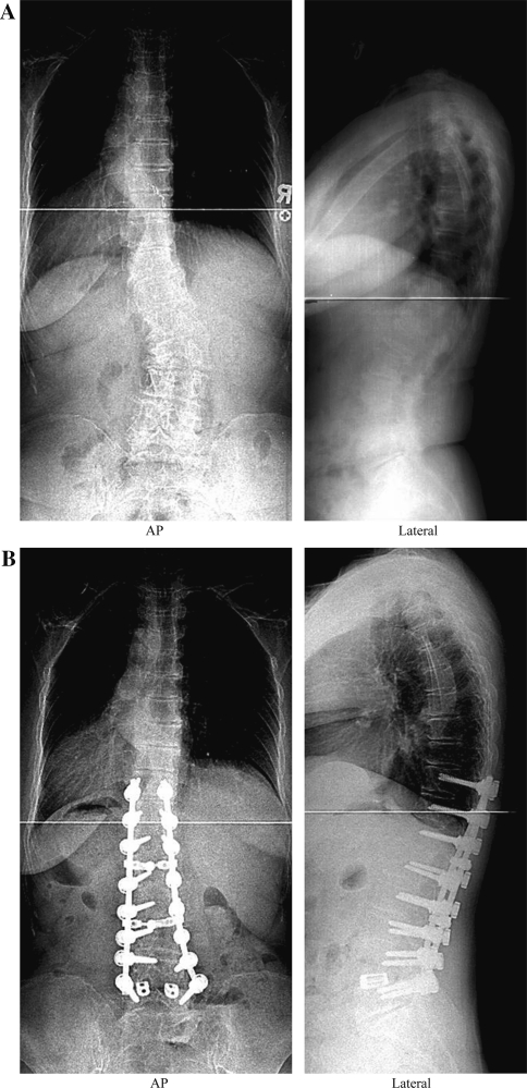
Fig. 2.
a Standing long cassette coronal and sagittal radiographs before surgery. The Cobb angle was 35° and lumbar lordosis was 54°. b Standing long cassette coronal and sagittal radiographs 2 years after long fusion and instrumentation with additional posterior interbody fusion at L4-5. The Cobb angle improved from 35 to 2°, and lumbar lordosis changed from 54 to 43°

