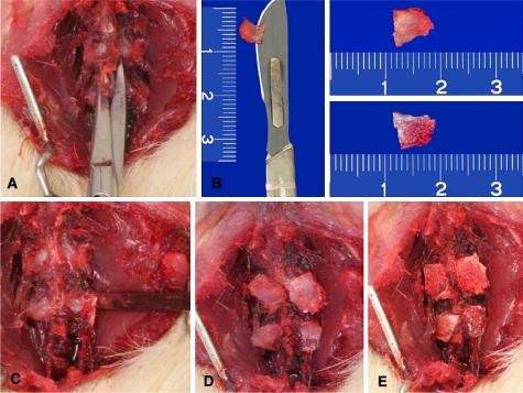
Fig. 1.
Photograph of the surgical stages of the experiment. a Exposure of the first two lumbar vertebrae and section at the base of the spinous processes. b The spinous processes were divided on the sagittal plane. Observe the surface of cortical bone (upper fragment) and cancellous bone (lower fragment). c Decortication of the posterior elements with a fine osteotome. d Apposition of the cancellous bone graft on the recipient bed. e Apposition of the cortical bone graft on the recipient bed

