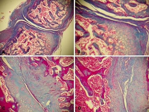
Fig. 5.
Photomicrographs of the interface of the non-decorticated group with a cancellous graft (group III). Observe the pattern of endochondral ossification. a Section from an animal killed 3 weeks after surgery, showing the formation of cartilage tissue, little contact between the surfaces in the interface and the formation of fibrous tissue around the interface (Masson trichrome, ×40). b Same section as in a at higher magnification (Masson trichrome, ×100) showing the partial filling of the interface with fibrous tissue and the presence of a cartilage mold that is being gradually replaced with bone tissue being formed at the periphery of the cartilage matrix. c Animal killed 6 weeks after surgery. Note the better contact between the surfaces in the interface and the gradual replacement of cartilage tissue with compact bone tissue (Masson trichrome, ×100). d Section from an animal killed 9 weeks after surgery. Observe the marked replacement of cartilage tissue with compact bone tissue, with a small amount of chondrocytes remaining at the periphery of the compact bone surfaces (Masson trichrome, ×100)

