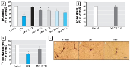
Figure 2.
Effect of fMLP on rat primary mesencephalic N/G cultures 8 days after treatment with vehicle (control), LPS (5 ng/mL) as a positive control, or different concentrations of fMLP. (A) DA neurotoxicity measured using the [3H]DA uptake assay; values are mean ± SD from four independent experiments in triplicate. (B) GABA neurotoxicity measured using the [3H]GABA uptake assay; values are mean ± SD from three independent experiments in triplicate. (C) Effect of fMLP (10−13 M) on dopaminergic neurons observed by immunocytochemistry staining with antibody against TH; DA neurotoxicity was measured by counting TH-IR neurons 8 days after treatment. Values are mean ± SD from three independent experiments in triplicate. (D) Representative images shown from three separate experiments for TH-IR neurons in a control culture and in cultures treated with LPS and fMLP. Bar = 50 μm.
*p < 0.05 compared with control.

