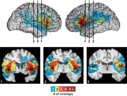
Fig. 2.
Lesion overlap in the insular cortex lesion group, in views of the right and left lateral surfaces, and coronal slices at three points moving anterior (1) to posterior (3) within the insular–somatosensory region. The colour bar indicates the number of overlapping cases at each voxel. Whilst all cases had unilateral lesions, the area of damage in the right- and left-sided cases was highly symmetrical. There is maximal lesion overlap across the group in the anterior insular cortex and somatosensory SII region, extending posteriorly into the inferior parietal cortex in some subjects.

