
Figure 4. Structural and functional characterization of Cy5-labeled his-ACBP.
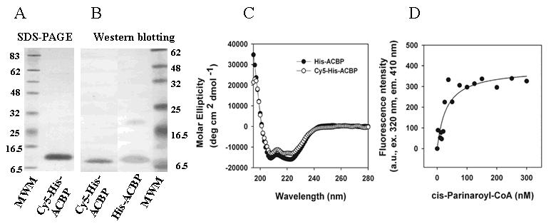
A. SDS-PAGE of Cy5-his-ACBP and his-ACBP. B. Western blotting of Cy5-his-ACBP and his-ACBP, 500ng of protein per lane. C. Far UV CD spectrum of his-ACBP and Cy5-his-ACBP. All CD studies were performed with 4μM protein. D. Titration and binding curve of Cy5-his-ACBP with cis-parinaroyl-CoA.