Figure 5.
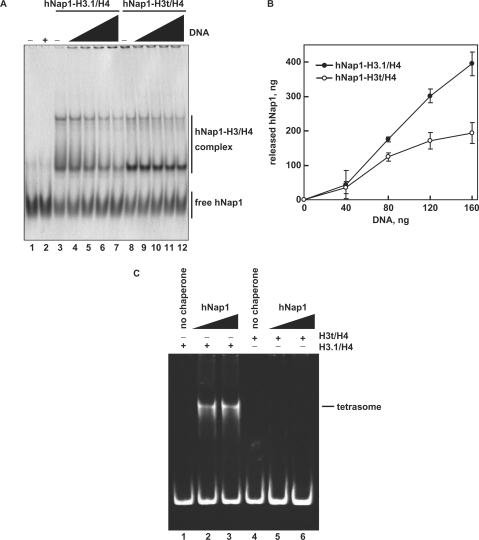
The H3/H4 deposition assay. (A) The deposition of H3.1/H4 or H3t/H4 from the hNap1-H3.1/H4 or hNap1-H3t/H4 complex onto DNA, analyzed by non-denaturing 5% PAGE with CBB staining. hNap1 (2.3 μg) was incubated without (lanes 1–2) or with H3.1/H4 (0.75 μg; lanes 3–7) or H3t/H4 (1.2 μg; lanes 8–12) to form the complex; about half of the hNap1 remained free under these conditions (lanes 3 and 8). After the incubation with supercoiled DNA, the samples were analyzed. The amounts of competitor DNA (ng) were 0 (lanes 3 and 8), 40 (lanes 4 and 9), 80 (lanes 5 and 10), 120 (lanes 6 and 11) and 160 (lanes 2, 7 and 12). (B) Graphic representation of hNap1 release. The amounts of hNap1 released from hNap1-H3.1/H4 (open circles) or hNap1-H3t/H4 complex (closed circles) are plotted as the averages of three independent experiments, performed as in (A), with the SD values. (C) Tetrasome-reconstitution with H3.1/H4 or H3t/H4 by hNap1. A total of 195 bp 5S DNA was incubated with hNap1 in combination with H3.1/H4 (8 ng/μl; lanes 1–3) or H3t/H4 (8 ng/μl; 4–6) (in the absence of H2A/H2B). The tetrasome formation was analyzed by non-denaturing 6% PAGE. The amounts of hNap1 (ng/μl) were 0 (lane 1), 45 (lane 2) and 182 (lane 3). The amounts of hNap2 (ng/μl) were 0 (lane 4), 43 (lane 5) and 171 (lane 6).

