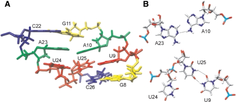
Figure 6.
Structural details within the asymmetric internal loop. (A) Side view of the average structure. Cytosines are colored in blue, adenines in green, uracils in red and guanines in yellow. (B) A10·A23 base pair and U9·U25·U24 base triple shown from the top. Dashed lines indicate hydrogen bonds.

