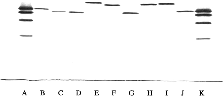
Fig. 1.
Isoelectric focusing of purified recombinant Hb mutants. Approximately 5 μg of each Hb was applied to the pH 6.0–8.0 range gel (Hb Resolve, Isolab) and electrophoresed for 30 min at 600 V and 45 min at 900 V at 10°C. The gel was stained with the JB2 stain (Perkin Elmer Wallach). The anode is at the top and the cathode is at the bottom. (Lane A) Standard hemoglobins A, F, S, and C from top to bottom. (Lane B) Hb V11I (β). (Lane C) Hb L3F(β). (Lane D) Hb L3F(β)/V11I(β). (Lane E) Hb P5E(β). (Lane F) Hb V1G(β)/P5E(β). (Lane G) Hb V1G(β)/P5A(β). (Lane H) Hb V1G(β)/P5E(β)/E7D(β). (Lane I) Hb V1G(β)/P5E(β)/E7D(β)/D21E(β)/E22D(β). (Lane J) HbA. (Lane K) Standard hemoglobins as in lane A.

