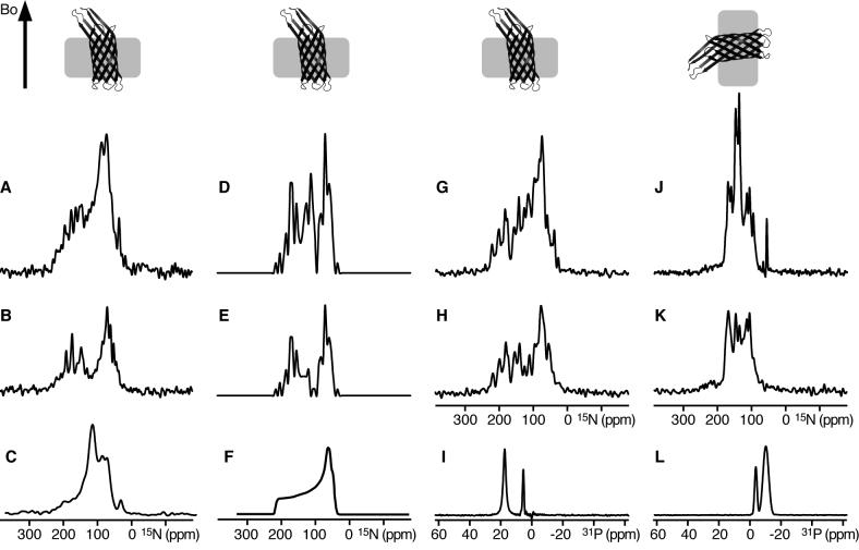
Figure 3.
Solid-state NMR spectra of uniformly 15N labeled OmpX in phospholipid bilayers. The corresponding orientations of OmpX membranes relative to the magnetic field (Bo) are shown at the top. (A, B) Supported DOPC/DOPG lipid bilayers oriented on glass slides with the membrane plane perpendicular to the magnetic field, in (A) H2O, or (B) D2O. (C) OmpX in unoriented 14-O-PC vesicles. (D, E) Spectra calculated from the crystal structure of OmpX rotated with the barrel axis tilted by 5° relative to the direction of the magnetic field (Bo), showing, (D) peaks from all sites, or (E) only peaks from β-strands but not from the loops. (F) Powder pattern calculated for static amide sites. (G-L) Lipid bicelles magnetically oriented with the membrane plane, (G-I) flipped perpendicular to the magnetic field by adding Yb3+, or (J-L) parallel to the magnetic field. Spectra were obtained (G, J) in H2O, or (H, K) in D2O. (I, L) 31P NMR spectra of the oriented bicelle lipids. The intensities of the experimental spectra were set to display a similar signal-to-noise level in the baseline. The intensities of the calculated spectra were set to match those of the experimental spectra.

