
Figure 4.
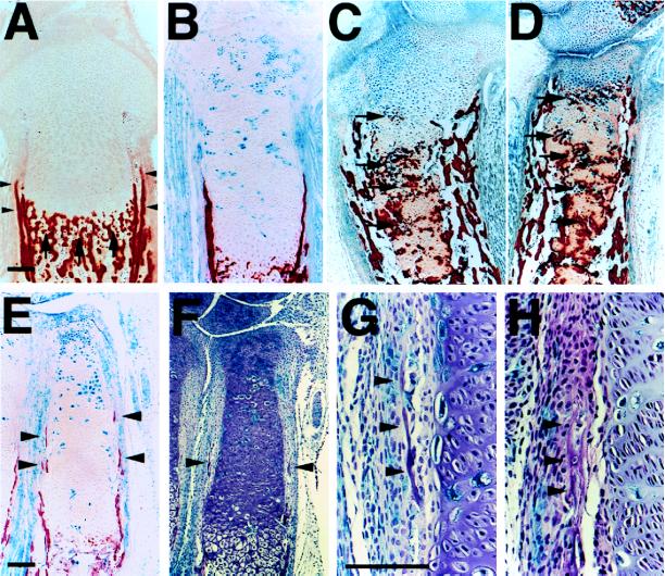
Ectopic mineralization of cartilaginous matrix and ectopic bone collar formation in chimeric bones. (A–D) Sections of the tibiae from d17.5 embryos were stained for β-galactosidase activity and also with alizarin red S for detection of mineralization. In the wild-type (A), mineralization of cartilaginous matrix occurs adjacent to late hypertrophic chondrocytes (arrows). No mineralization is observed adjacent to ectopic hypertrophic chondrocytes in the tibiae from chimeras with ≈60% contribution of PTH/PTHrP receptor (−/−) cells (B), whereas in chimeras with >90% contribution, matrix adjacent to ectopic hypertrophic chondrocytes in large clusters mineralize (C and D, arrows). (E–H) Sections of the radii from d17.5 chimeric embryos with ≈60% contribution of mutant cells were stained for β-galactosidase activity and also stained with alizarin red S (E) or H&E (F–H). In the wild-type (A), bone collars are formed in the perichondrium abutting prehypertrophic and hypertrophic chondrocytes (arrowheads). No ectopic bone collar formation is observed in the tibiae of chimeras with ≈60% contribution (B). However, in smaller bones such as the radii of the same chimeras, ectopic bone collars are formed (E and F, arrowheads). At higher magnification, ectopic bone collars are seen near clusters of ectopic hypertrophic chondrocytes; ectopic bone collars include both wild-type and PTH/PTHrP receptor (−/−) osteoblasts (stained blue) (G and H, arrowheads). In the tibiae of chimeras with >90% contribution, there is intensive ectopic bone collar formation (C and D). (Bar = 100 μm.)