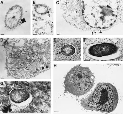
Figure 5.
Electron microscopic visualization of CFTR interacting with P. aeruginosa. (A and B) P. aeruginosa cells are seen with accumulations of CFTR at a single point on the bacterial surface (arrow). (Bar = 0.1 μM; gold particles are 30 nm.) (C) Epithelial cells not ingesting P. aeruginosa showed primarily intracytoplasmic CFTR (open arrows) and membrane CFTR usually only bound to one or two gold particles (closed arrows) and, on a rare occasion, small aggregations of gold particles (closed arrowhead). (Bar = 0.5 μM; gold particles are 30 nm.) (D) Control where the primary antibody to CFTR was omitted showing epithelial cell with internalized P. aeruginosa bacteria (indicated by “b” on figure). (Bar = 0.1 μM.) (E–G) Epithelial cells not treated with methanol (used to permeabilize the cells in A–D for intracytoplasmic antibody reactions) showed P. aeruginosa in intracytoplasmic enclosures with only a portion of the bacterial cell surface attached to the vesicle membrane (arrow), similar to the accumulation of CFTR on the bacterial surface (A and B). (Bar = 0.1 μM in E; 0.5 μM in F and G.) (H) Epithelial cells homozygous for the ΔF508 CFTR mutation could not be seen ingesting P. aeruginosa. (Bar = 1.0 μM.)

