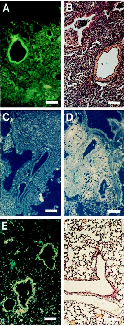
Figure 6.

Immunofluorescence of CFTR in lung sections of mice infected with P. aeruginosa. (Bars = 100 μM.) (A) CFTR visualized by mAb CF3 and fluoresceinated secondary antibody in the airway epithelium of mice infected with P. aeruginosa 24 hr previously. (B) Hematoxylin and eosin stain of a lung section from a neonatal mouse infected with P. aeruginosa. (C) Visualization of CFTR by mAb CF3 and fluoresceinated secondary antibody in the airway epithelium of mice infected with P. aeruginosa 24 hr previously was inhibited if the mAb was first incubated with the first extracellular domain synthetic peptide (amino acids 103–117 of CFTR). (D) CFTR could not be visualized by mAb CF3 and fluoresceinated secondary antibody in the airway epithelium of mice infected 24 hr previously with P. aeruginosa along with a synthetic peptide corresponding to the first predicted extracellular domain of CFTR. This synthetic peptide inhibited CFTR-mediated epithelial cell internalization of P. aeruginosa and the subsequent clearance of the bacterium from the lung (see Fig. 4). (E) Staining of uninfected mouse lung section with mAb CF3 and fluoresceinated secondary antibody. Little to no CFTR is seen in the epithelium. (F) Hematoxylin and eosin stain of a lung section from an uninfected neonatal mouse.
