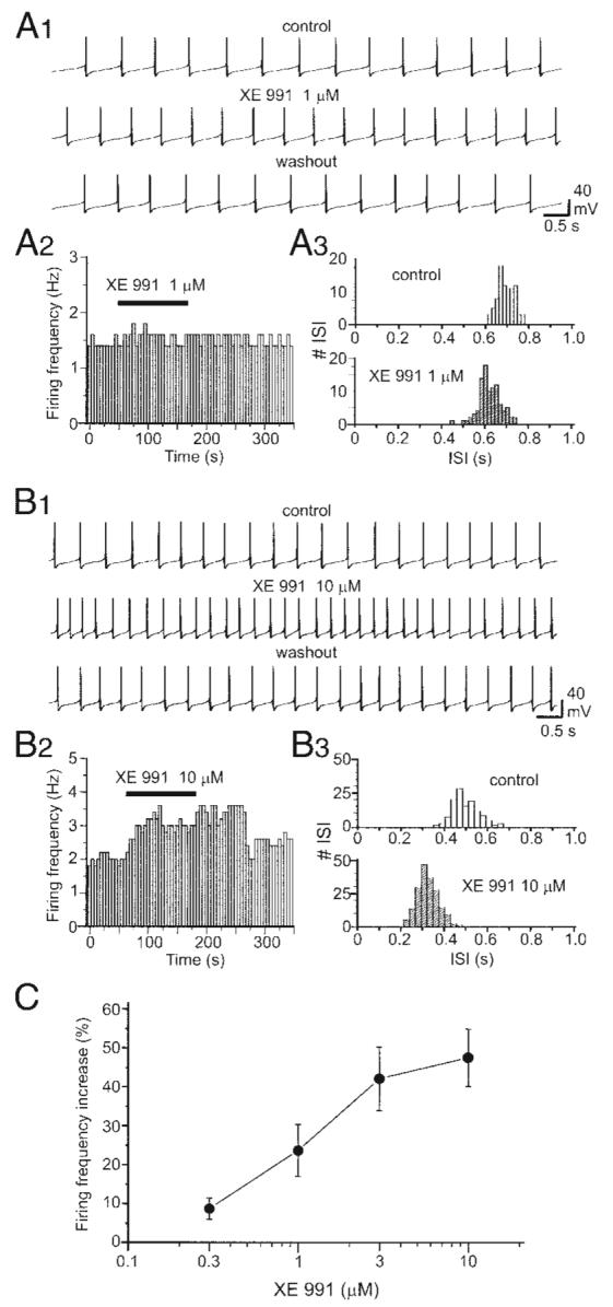
FIG. 5.

Blockade of IM increases the firing frequency of DA VTA neurons. A1: current-clamp recording of spontaneous firing from a typical DA VTA neuron before (top), during (middle), and after application of 1 μM XE991 (bottom). A2, left: time course of the effect of 1 μM XE991 from the same neuron in A1. Each vertical bar represents average firing frequency over a 5-s-long interval. Right: ISI histogram before (white columns) and after treatment with 1 μM XE991 (striped columns) (bin width, 17 ms). B1: current-clamp recording of spontaneous firing from a typical DA VTA neuron before (top), during (middle), and after application of 10 μM XE991 (bottom). B2: time course of the effect of 10 μM XE991 from the same neuron in B1.B3: ISI histogram before (top) and after treatment with 10 μM XE991 (bottom) (bin width, 35 ms). C: concentration–response curve for XE991-induced firing rate increase in the population of DA VTA neurons tested (0.3 μM, n = 7; 1 μM, n = 7; 3 μM, n = 6; 10 μM, n = 8).
