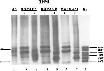
Figure 4.
Western blots of native and dephosphorylated tau from the DDPAC and Montreal kindreds. Insoluble fractions obtained from the frontal cortex of two different brains of a DDPAC kindred and a brain from the Montreal kindred were probed with the two phosphorylation independent mAbs T14 and T46. Dephosphorylation reveals two major and one minor band that align with all recombinant 4Rtau isoforms (lanes 3, 5, and 7). The major bands align with 0N4Rtau and 1N4Rtau isoforms, and the minor band aligns with the 2N4Rtau isoform. Molecular weight standards are shown in the left margin, and the positions of each of the nonphosphorylated recombinant tau isoforms are labeled. Lane 1 shows PHFtau from the brain of a patient with AD. Lanes 2 and 4 show native tau, and lanes 3 and 5 show dephosphorylated tau from two different DDPAC brains. Lanes 6 and 7 show native and dephosphorylated tau from a brain of the Montreal kindred. Abbreviations are as in Fig. 3.

