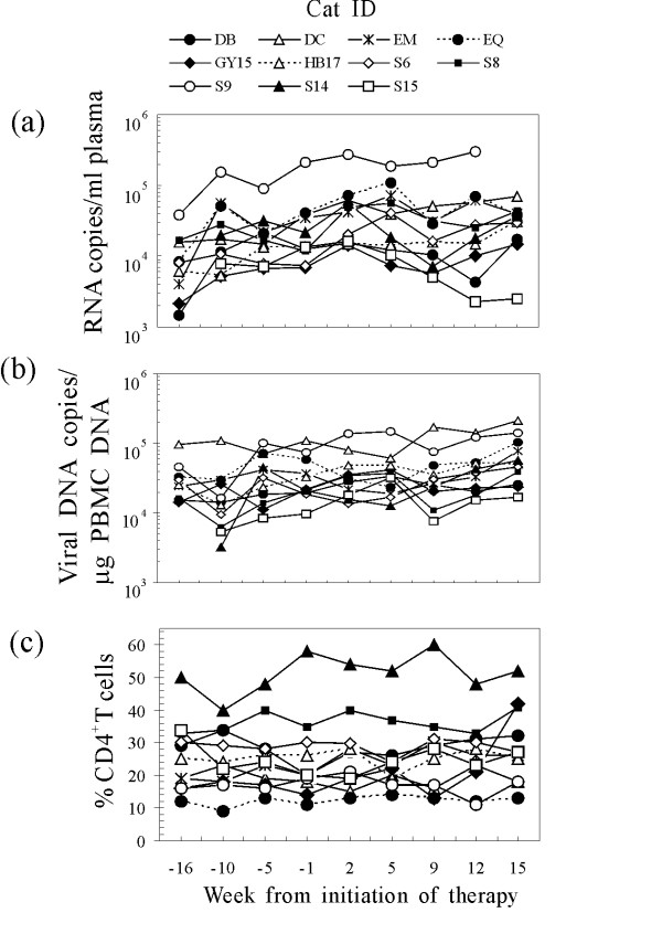
Figure 1.
Plasma viremia (a), proviral load in the PBMCs (b), and circulating CD4+ T lymphocyte percentages (c) of the study cats at the times indicated relative to the first FIV-MDC inoculum. CD4+ T cells were monitored by flow cytometry in peripheral blood by direct staining with anti-feline CD4-PE (clone vpg34, AbD Serotec, Raleigh, NC) for 30 min as previously described (8). Symbols represent individual animals. Arrows indicate the times of FIV-MDC inoculation.

