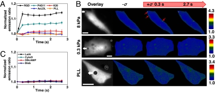
Fig. 2.
Src activation depends on stress probe specificity, substrate rigidity, intact F-actin, and prestress. (A) Probe specificity on Src activation. Mechanical stimulation via the magnetic bead coated with RGD or anti-β1-activating antibody (P4G11), induced Src activation in the cytoplasm but not anti-β1-nonactivating antibody (K20), AcLDL (binds scavenger receptors), or PLL (strong nonspecific surface binding) (Fig. S5). RGD, n = 12 cells; P4G11, n = 4 cells; K20, n = 3 cells; AcLDL, n = 4 cells; PLL, n = 3 cells. Error bars represent SEM. (B) Mechanical stimulation of cells plated on soft (0.3 kPa, n = 10 cells) polyacrylamide gel substrate did not induce Src activation, whereas the cell on relatively hard (8 kPa, n = 4 cells) substrate induced strong Src activation (red arrows). Cells on PLL substrate (n = 8 cells) that do not form basal focal adhesions and stress fibers (12) did not activate Src in response to mechanical stress. White arrows indicate magnetic bead movement direction (stress = 17.5 Pa). (Scale bars, 10 μm.) (C) Preincubating cells with CytoD (1 μg/ml for 15 min; n = 5 cells), LatA (1 μM for 15 min; n = 4 cells) to disrupt actin microfilaments, Bleb (50 μM for 20 min; n = 5 cells) to inhibit myosin II, or DBcAMP (1 mM for 15 min; n = 4 cells) to relax the cell, prevented stress-induced Src activation (Fig. S6).

