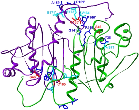
Fig. 3.
Location of the residues found changed in AhpC in cytoplasmic thiol redox suppressor strains. Shown is an AhpC dimer (subunits in green and purple) in the structure of the C46S mutant (Protein Data Bank ID code 1N8J). C165 and C46 (replaced with serine in this structure), are shown in red. Residues found mutated in the ΔtrxB Δgor supp strain are in blue (3, 4). The ahpC* mutation results in the addition of a phenylalanine residue at Y38. Residues found mutated in the ΔgshA ΔtrxB supp strain are in cyan.

