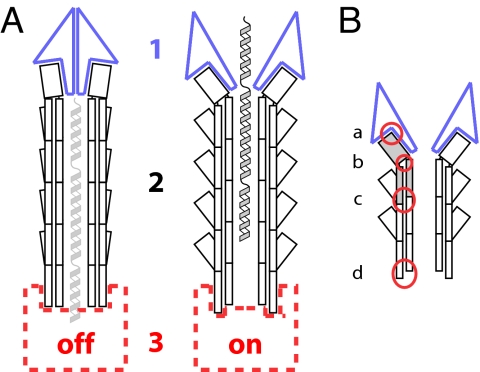
Fig. 4.
Schematic illustrations of possible different states of the T3SS needle complex and sites of action for needle mutants. (A) Changes in three distinct regions of the T3SS may be involved in the regulation of secretion after host-cell sensing: the tip complex (1, blue), the needle itself (2, black), and the basal apparatus (3, red). Secretion substrates are shown in the needle channel in a partially unfolded state (light gray and gray). (B) Mutations of the needle protein can alter secretion patterns by disruption of four different interfaces (red circles): needle subunit to tip complex (a), within the needle subunit, i.e., intramolecular (b), between the needle subunits, i.e., intermolecular (c), and needle subunit to base (d).

