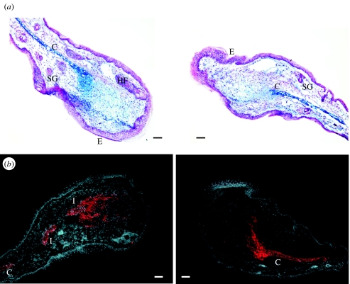
Figure 2.
Transverse sections through an MRL/MpJ ear 21 days post-biopsy punch wounding. The histological organization is blastema-like in structure. (a) shows an ear section stained with Alcian Blue and fast red. The cut end of the cartilage (C) can be seen clearly. Glycosaminoglycan deposits in the mesenchyme are stained blue. The apical epithelium (E) extends away from the cut cartilage. (b) shows a similar ear section stained with anti-Aggrecan-TRITC, a cartilage precursor molecule. The section is counterstained with the nuclear dye, DAPI. Note the formation of cartilage islands (I) in the mesenchyme and the infiltration of Aggrecan into the mesenchyme. HF, hair follicle; SG, sebaceous gland. Scale bar, 100 μm.

