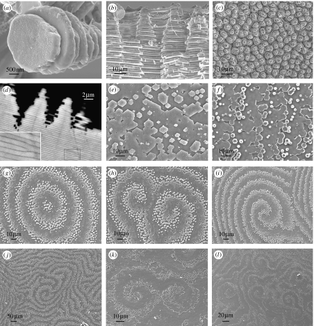
Figure 1.
Nacre morphology. Scanning electron micrographs of (a–d) gastropod nacre: Calliostoma zyziphynus showing (a) a tower of tablets and (b) a fractured transverse section. (c) Bolma rugosa displaying towers of tablets and (d) Gibbula pennanti back scattered electron scanning of a transverse section: (e–l) bivalve nacre; (e) Anodonta cygnea, (f) Atrina pectinata displaying growth fronts made up of tablets, (g–j) Pteria avicula and (k) and (l) Pteria hirundo, showing target, spiral and labyrinthine patterns at the mesoscale, respectively.

