Figure 3.
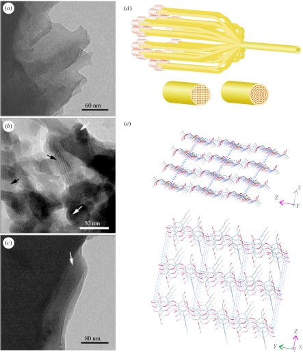
Chitin in nacre. Cryo-transmission electron micrographs of a vitrified suspension of fixed and demineralized nacreous shell organic matrix fragments from Atrina. (a) A matrix fragment showing the homogenous texture and layered structure. (b) An aggregation of small fragments aligned such that, in some areas, lattice images are visible (black arrows). White arrows show net-like structures. (c) A fragment of the organic matrix after the suspension was refluxed in 1 M NaOH to remove the protein. The homogenous texture and layered structure are still preserved, as is the lattice image in part of this fragment (arrow). (a–c) are taken from figure 1 in Levi-Kalisman et al. (2001). (d) Sketch of the putative chitin crystallization process showing alternative possibilities for the alignment of the crystal planes dictated by the arrangement of the rosettes extruding the polymer chains. (e) Crystal structure of β-chitin (Dweltz et al. 1968).

