Fig. 9.
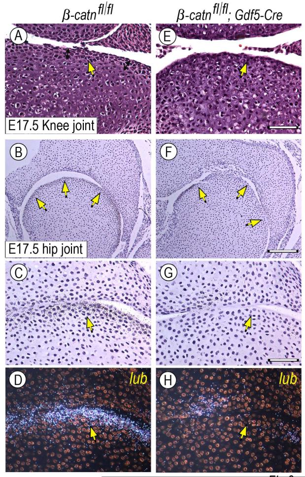
Defects in superficial flat cell layer and gene expression patterns in conditional Gdf5-Cre β-catenin-deficient joints. E17.5 knee and hip joints from control (β-catn fl/fl) and β-catenin-deficient (β-catn fl/fl/Gdf5-Cre) mouse embryo littermates were analyzed by standard histology and for expression of lubricin. (A-D) Control joints display the high cell density flat cell layers along the joint perimeter (A-C, arrows) and lubricin (lub) transcripts (D). (E-H) Instead, the superficial high-density layers are much less obvious in mutant joints (E-G, arrows) and lubricin expression is much reduced (H). Bar for A and E, 50 μm; bar for B and F, 175 μm; bar for C-D and G-H, 60 μm.

