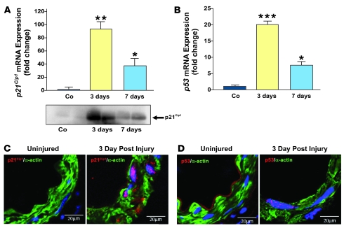
Figure 2. p21Cip1 and p53 are induced after vascular injury.
Vascular wire injury was performed on p21+/+ mice, and femoral arteries were harvested 3 and 7 days following injury. (A) Upper panel: Total RNA was isolated from femoral arteries, and endogenous p21Cip1 mRNA was quantified by quantitative PCR and normalized to levels of 18S RNA. Levels of p21Cip1 mRNA at 3 (yellow bar) and 7 days (blue bar) are expressed relative to that measured in uninjured control (Co) arteries (n = 3; *P < 0.05 versus Co; **P < 0.01 versus Co). Lower panel: Western blot analysis of p21Cip1 levels in femoral arteries at 3 and 7 days after injury compared with uninjured arteries. (B) Levels of p53 mRNA at 3 (yellow bar) and 7 days (blue bar) are expressed relative to that measured in uninjured arteries (n = 3; *P < 0.05 versus Co; ***P < 0.001 versus Co). p21Cip1 (C) and p53 (D) staining were detected by confocal microscopy in VSMCs arteries 3 days after injury. VSMCs were identified by smooth muscle α-actin (green), p21Cip1 (C, red), p53 (D, red), and nuclear counterstaining by DAPI (blue). p21Cip1 and p53 appear pink due to the overlay with the blue DAPI staining.

