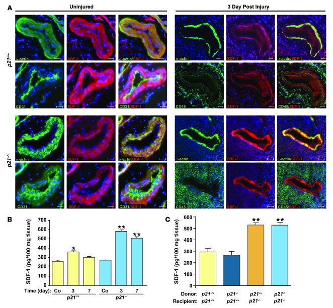
Figure 6. SDF-1 is localized to the arterial media, and SDF-1 tissue levels are increased in p21–/– mice receiving p21+/+ BM after arterial injury.
(A) SDF-1 staining in uninjured arteries (left panels) and in arteries 3 days after injury (right panels) from p21+/+ (upper panels) and p21–/– (lower panels) mice. VSMCs were identified by smooth muscle α-actin (in green), endothelial cells by CD31 (in green), and inflammatory cells by CD45 (in green). SDF-1 staining is represented in red, and nuclei were counterstained by DAPI (blue). For uninjured and injured groups, left and middle columns represent individual staining; right columns are merged images. Scale bars: 40 μm. (B) Increased arterial tissue SDF-1 levels were observed in p21–/– (blue bars) compared with p21+/+ mice (yellow bars) 3 and 7 days after injury (n = 3; **P < 0.01 versus p21+/+ at same time point). Increased SDF-1 levels were also observed in p21+/+ mice 3 days after injury compared with uninjured p21+/+ mice (n = 3; *P < 0.05). (C) Arterial SDF-1 levels measured 7 days after injury were increased in p21–/– mice that received either p21+/+ (orange bar) or p21–/– (light blue bar) BM compared with p21+/+ mice that received either p21+/+ (yellow bar) or p21–/– (dark blue bar) BM, respectively (n = 5; **P < 0.01).

