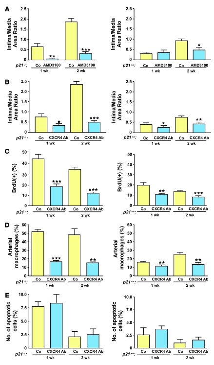
Figure 7. SDF-1 inhibition prevents excessive proliferation during vascular wound repair.
(A) Reduced vascular lesions in p21–/– (left) and p21+/+ arteries (right) after AMD3100 treatment compared with saline-treated animals (Co) 1 and 2 weeks after vascular injury (left, n = 5; right, n = 10). (B) Reduced vascular lesions in p21–/– and p21+/+ arteries with anti-CXCR4–blocking antibody compared with mice treated with IgG control (left, n = 5; right, n = 16). (C) Anti-CXCR4 blocking antibody decreased cellular proliferation as assessed by BrdU incorporation at 1 and 2 weeks after vascular injury in p21–/– and p21+/+ arteries (left, n = 5; right, n = 5). (D) Anti-CXCR4 blocking antibody reduced the number of local arterial macrophages in p21–/– and p21+/+ arteries at 1 and 2 weeks after vascular injury (left, n = 5; right, n = 5). (E) The number of local apoptotic TUNEL-positive cells after vascular injury was unchanged after treatment with anti-CXCR4 blocking antibody. *P < 0.05, **P < 0.01, and ***P < 0.001 versus Co at same time point.

