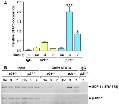
Figure 9. STAT3-dependent SDF-1 signaling pathway is impaired in the p21–/– arterial wall.
ChIP assays on p21+/+ and p21–/– femoral arteries using anti-STAT3 antibodies were performed before femoral wire injury (Co) and 3 and 7 days after injury. (A) Quantification of STAT3 occupancy within the SDF-1 promoter region in p21+/+ and p21–/– femoral arteries (n = 5; *P < 0.05, ***P < 0.001 versus same time point for p21+/+). As a control, chromatin from p21–/– arteries 3 days after injury was immunoprecipitated with IgG. (B) PCR amplification of the SDF-1 (–572/–372 bp) and β-actin promoters after ChIP with IgG or anti-STAT3 antibodies following vascular injury.

