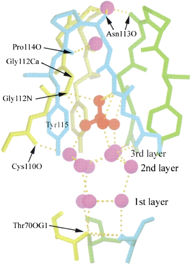
Fig. 6.

The trap for a sulfate ion at the open interface of the RNase A minor trimer. The sulfate ion and the water molecules are in red and purple, respectively. The protein chains from the three subunits of the minor trimer are in cyan, green, and yellow, respectively. The protein atoms and residues that hydrogen bond with the water molecules and sulfate ion are indicated. There is an intricate hydrogen bond network in the trap. The waters in the trap are aligned in three layers as labeled. The structure is viewed perpendicular to the threefold axis of the minor trimer. For clarity, the sidechains of Cys 110, Glu 111, and Asn 113 are omitted. The figure was created using SETOR (Evans 1993).
