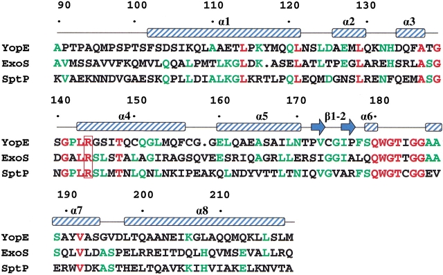
Fig. 2.
Structure-based sequence alignment of the bacterial GAP domains. Residues that are identical in all three sequences are shown in red; residues identical in two out of three sequences, in green. The critical arginine residue is enclosed by a red box. The positions of α-helices and β-strands are indicated above the sequence. Residues are numbered according to the full-length YopE sequence.

