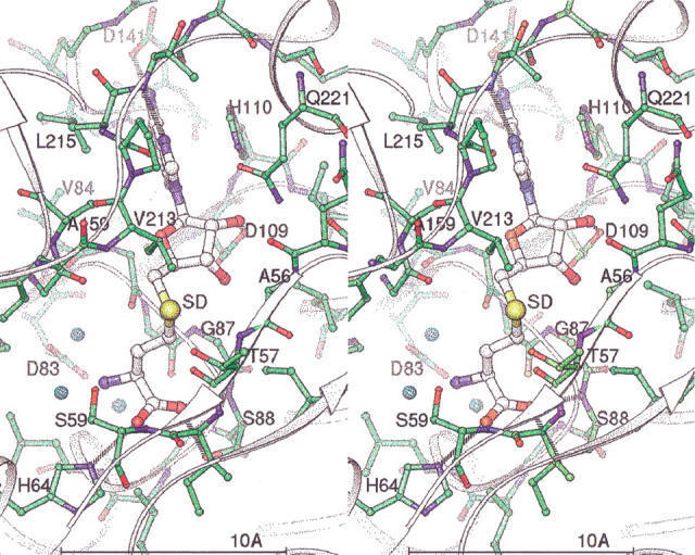
Fig. 3.

Stereo view of the AdoHcy binding site. Key residues and atoms are labeled. Atoms are colored by type, with green carbon/bonds for the protein and white carbon/larger bonds for AdoHcy. Three tightly bound waters are shown as small cyan spheres. Hydrogen bonds between the protein and AdoHcy are shown as dashed lines.
