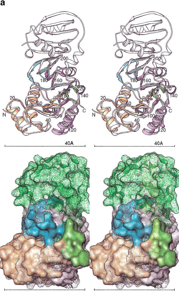
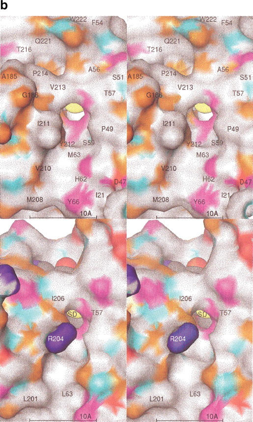
Fig. 5.


(a) Superposition of HPIMT and TMPIMT structures. (Top) Stereo ribbons diagrams. The human structure is colored by its sequence domains: yellow-orange is the N-terminal, lavender is the conserved MT core, cyan are the sheets with the unique topology in PIMTs, and green is the divergent C-terminal region. The termini and every 20 residues are labeled. The AdoHcy molecule is colored by atom type. The superposed TMPIMT backbone is white and its AdoHcy gray. (Bottom) Stereo molecular surfaces. The human structure is solid and colored as above. The TMPIMT surface is shown as a mesh, colored white for the common catalytic domain and green for its divergent and unique C-terminal domain. (b) PIMT comparison of substrate-binding surface. The molecular surfaces are colored by chemical property: white, hydrophobic; red, negative; blue, positive; orange, H-bond acceptor; cyan, H-bond donor; and magenta, polar. Key residues are labeled. The AdoHcy molecule is shown as spheres colored by atom type. (Top) HPIMT. (Bottom) TMPIMT.
