Fig. 7.

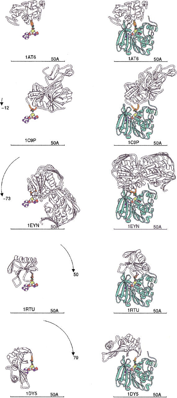
IsoAsp-containing protein binding models. Each PDB file containing isoAsp is shown as a white ribbon with the five residues centered on isoAsp colored orange, and the isoAsp is shown as a stick figure. Each protein is labeled by its PDB code. The AdoMet as bound to human PIMT provides the reference point, shown as spheres colored by atom type. (Left) PDB proteins aligned to superpose their isoAsp residue with the isoAsp modeled into the HPIMT site (Fig. 6 ▶). The rotation to be applied is shown on the arc. (Right) PDB isoAsp proteins docked into HPIMT by a single rotation about the isoAsp loop as explained in the text. HPIMT is shown as a light green ribbon. PDB codes 1AT6: hen egg white lysozyme; 1C9P: porcine trypsin; 1EYN: enoylpyruvate transferase; 1RTU: Ustilago sphaerogena ribonuclease U2; 1DY5: bovine pancreatic ribonuclease.
