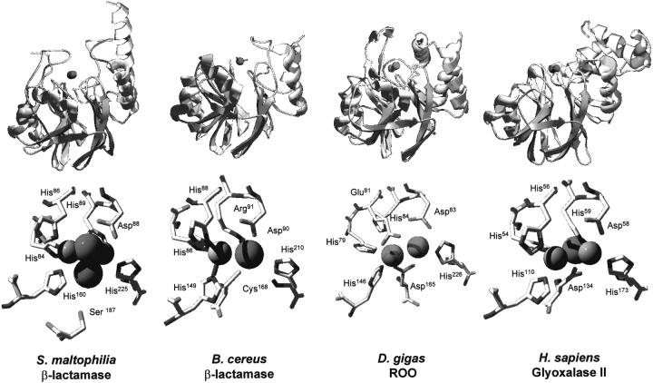
Fig. 1.
Comparison of the three-dimensional structures of proteins with the αβ/βα fold of class B β-lactamases. (Top) Ribbon representation of the regions of the studied proteins comprising the class B β-lactamase fold; (bottom) structures of bimetallic centers from the same proteins. The representative structures here depicted are the zinc β-lactamases from Bacillus cereus (2bc2) and S. maltophilia (1sml), and the structural domains of D. gigas rubredoxin:oxygen oxidoreductase (1e5d) and H. sapiens glyoxalase II (1qh5) that also have a αβ/βα lactamase-like fold. The protein structures were superimposed using Swiss-PdbViewer (Guex and Peitsch 1997) and are represented in identical configurations. For definition, metal site position 1 is on the left and position 2 on the right. Metal-ligating water molecules are represented as spheres. Rendering was done with POV-ray.

