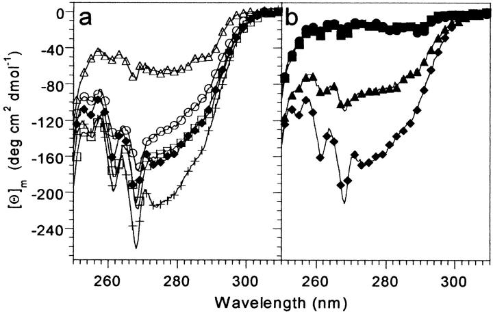
Fig. 3.
Near-UV CD spectra of the VHP subdomain mutants at 4°C. (a) Single leucine point mutants. (b) Double leucine mutants. Samples were 100 to 225 μM protein, in 50 mM phosphate buffer, pH 7.0. The M53L spectrum is shown in both (a) and (b) for comparison. HP36 (+); M53L (filled diamonds); F47L (open circles); F51L (open squares); F58L (open triangles); F47,51L (filled triangles); F47,58L (filled squares); F51,58L (filled circles).

