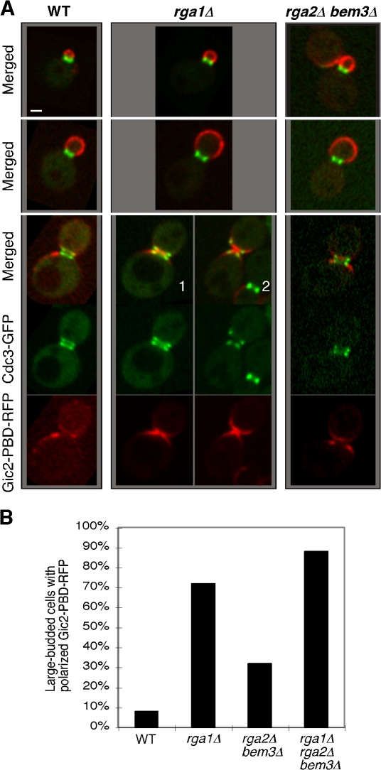
Figure 3.
Deletion of RGA1 causes an elevated level of GTP-Cdc42 at the cell division site. (A) Cells of YZT292 (WT), YZT293 (rga1Δ), and YZT294 (rga2Δ bem3Δ) carrying integrated CDC3-GFP and GIC2-PBD-RFP were imaged by two-color light microscopy. Single representative GFP and RFP images from a stack of z sections for each cell were selected to show the localization patterns of Cdc3-GFP and Gic2-PBD-RFP with high resolution. Bar, 1 μm. (B) Quantitation of large-budded cells with neck-localized Gic2-PBD-RFP. Cells of YZT295 (rga1Δ rga2Δ bem3Δ) and other strains as in A were used and only large-budded cells with a clear septum (n = 50 for each strain) were scored.

