
Fig. 1.
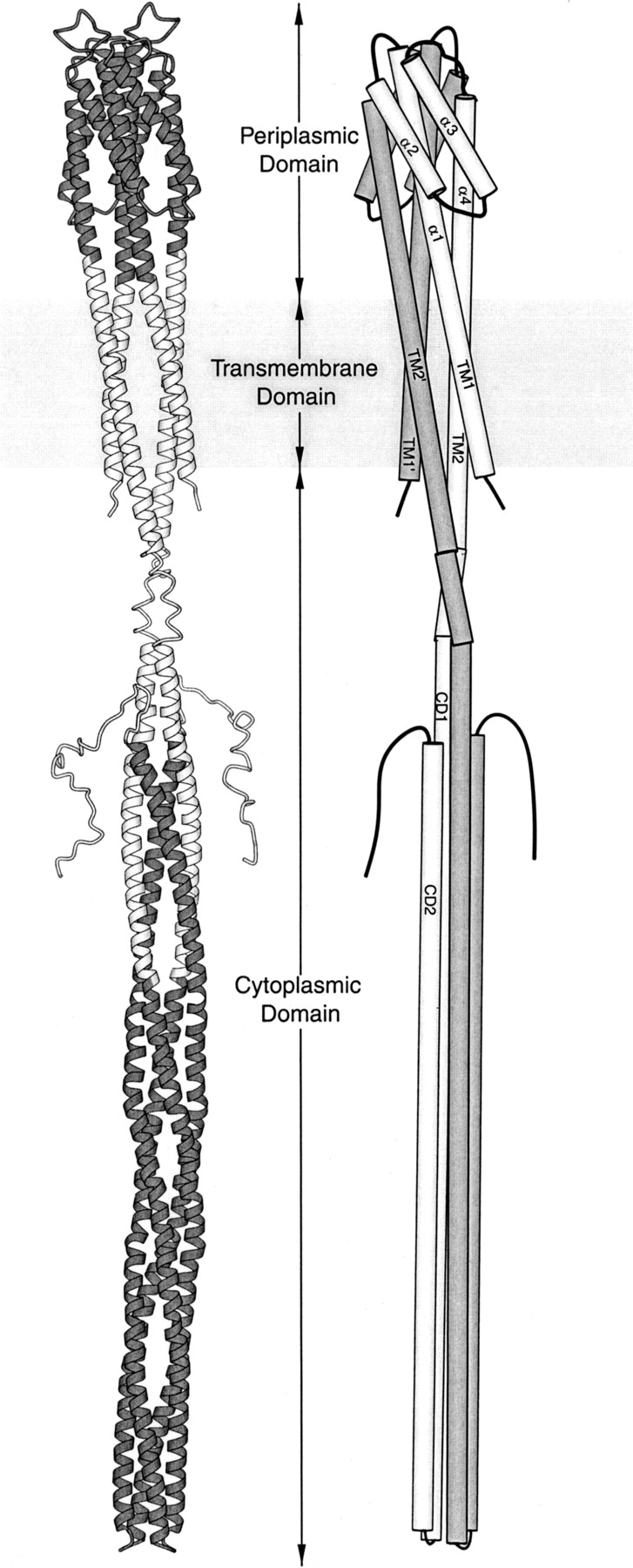
Models of chemoreceptor structure. In a ribbon diagram (left) of a homodimeric bacterial chemoreceptor, the portions of the model based on X-ray crystallography of receptor fragments are shaded. In a schematic diagram (right), helices are labeled, subunits are differentially shaded, and helix super-coiling is omitted. Each ∼60-kD subunit begins on the cytoplasmic side of the membrane, crosses the membrane as transmembrane helix 1 (TM1), becomes helix α1 of the periplasmic domain, turns to make the membrane-distal four-helix bundle, extends to the membrane as helix α4, crosses the membrane as transmembrane helix 2 (TM2), extends into the cytoplasm, and makes a membrane-distal turn to create the extended helical hairpin of cytoplasmic helices 1 and 2 (CD1 and CD2). The ligand-binding site is at the membrane-distal end of the periplasmic domain, and the kinase-interaction site is at the membrane-distal end of the cytoplasmic domain. This figure was made using Molscript (Kraulis 1991).