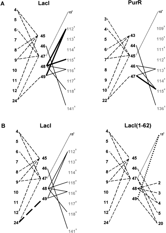
Fig. 8.
Maps of N-linker contacts for LacI, PurR, and LacI(1–62). N-linker residues of the protein monomers are indicated by large black numbers, as are sites on the HTH of the same monomer. Sites in the core domains are shown as large gray numbers, and DNA positions as small black numbers. The prime symbol indicates a residue of the partner monomer or a base of the cognate strand. Solid lines depict interactions to the partner core domain, dashed lines represent intrasubunit HTH contacts, and dotted lines are interactions with DNA. Bold lines (solid, dash, or dot) indicate an additional interaction in one of a pair of proteins. (A) LacI versus PurR. Homologous sites align horizontally. For example, LacI 5 is homologous to PurR site 3. (B) LacI versus LacI(1–62). Additional N-linker contacts to the HTH of the same monomer are indicated on the right of the diagram for LacI(1–62); these occur in place of full-length LacI N-linker interactions with the partner core domain.

