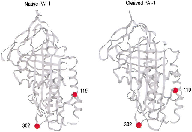
Fig. 1.
Structures of native and cleaved PAI-1 showing the two positions of fluorescent label incorporation (residues 119 and 302). Ribbon diagrams were drawn using Rasmol with coordinates for native PAI-1 (left) from structure 1b3k (Sharp et al. 1999) and for cleaved PAI-1 (right) from 9pai (Aertgeerts et al. 1995). The reactive center loop is at the top. The location of the proteinase in the α1PI:trypsin complex is at the bottom of the structure, impinging upon residue 314, which is the equivalent of residue 302 in PAI-1.

