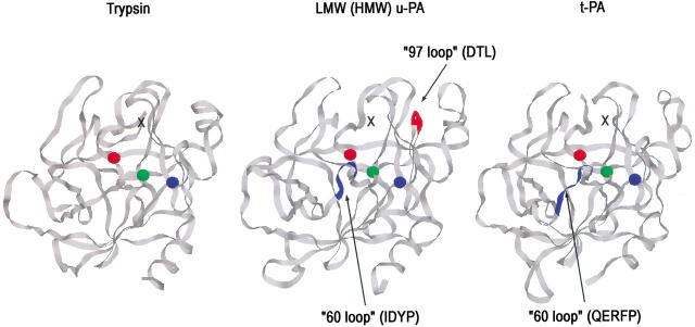
Fig. 7.
View of the putative contact surfaces of trypsin, LMW- and HMW u-PA, and t-PA in covalent complexes with PAI-1, looking from the serpin toward the proteinase catalytic domain. The black crosses mark the possible location of residue 302 of PAI-1 in each complex based on the location of the structurally equivalent residue 314 of α1PI in the covalent α1PI:trypsin complex. Residues marked as red, green, and blue spheres are the catalytic triad serine 195, histidine 57, and aspartate 102, respectively. The 60 and 97 insertion loops are shown as blue or red ribbons. The structures were drawn with Rasmol 2.7 using the pdb coordinate files 1SMF for trypsin (Li et al. 1994), 1LMW for u-PA (Spraggon et al. 1995), and 1RTF for t-PA (Lamba et al. 1996).

