
Fig. 5.
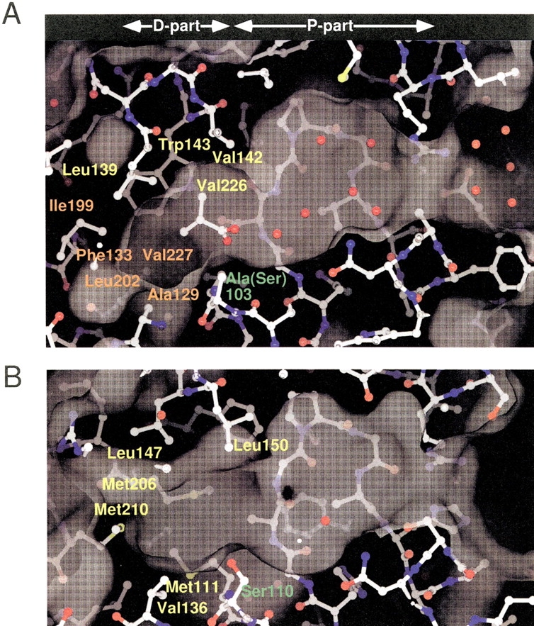
Molecular surfaces of the substrate-binding pockets of CumD (A) and RHA1 BphD (B), calculated using the SPOCK program with probe radius of 1.4 Å. Waters are shown as red spheres. The catalytic serine or mutated alanine residues are labeled in green. The residues involved in the recognition of the isopropyl group of ISB300 and in the formation of the deeper space of the D-part are labeled in yellow and orange, respectively. The residues involved in the formation of the surface of the D-part of RHA1 BphD are labeled in yellow.