
Fig. 6.
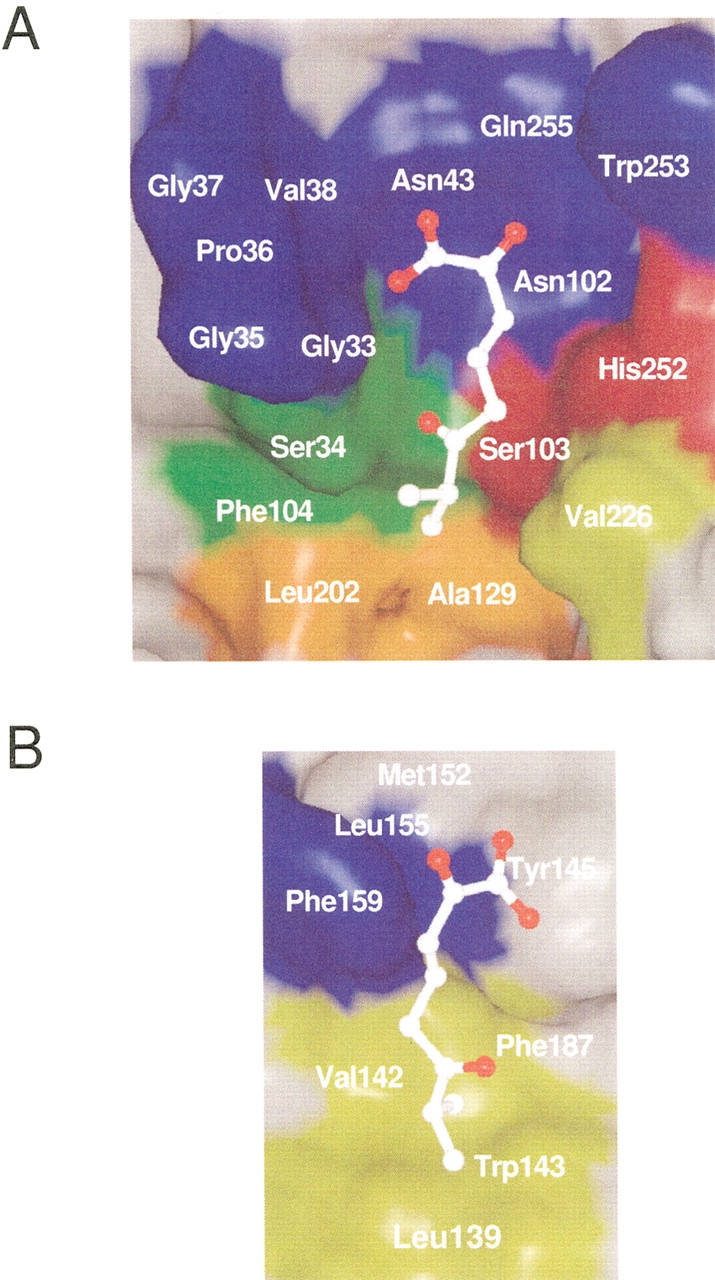
Modeled substrate in the substrate-binding pocket. The inner surface of the core-domain side (A) and that of the lid-domain side (B) are shown. The side-chain of Ser103 was modeled and energy-minimized with the substrate (see Materials and Methods). The modeled substrate is shown with a ball-and-stick model. The inner surface formed by the residues involved in the catalysis, in the recognition of the carboxylate group and isopropyl group of isobutyric acid, and in the formation of the deeper space of the D-part are colored in red, green, yellow, and orange, respectively. Other conserved residues, which are shown in pink in Fig. 2 ▶, are shown in blue.